Authors: Shadab Ahamed, Yixi Xu, Ingrid Bloise, Joo H. O, Carlos F. Uribe, Rahul Dodhia, Juan L. Ferres, Arman Rahmim
Published on: March 11, 2024
Impact Score: 7.2
Arxiv code: Arxiv:2403.07105
Summary
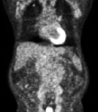
- What is new: This study introduces an approach for automated slice classification in medical imaging, specifically for identifying slices potentially containing tumors in lymphoma PET/CT images, utilizing a novel training configuration.
- Why this is important: The challenge of quickly and accurately identifying image slices that potentially contain tumors from vast PET/CT image datasets, to improve the efficiency of medical image segmentation and diagnostic processes.
- What the research proposes: A method using the ResNet-18 network trained on axial slices from PET/CT images with a combination of patient-level split datasets, input types, and training strategies to accurately classify slices containing tumors.
- Results: The best model was trained with patient-level split data and CAG strategy on PET slices only, showing superior performance in most metrics, particularly in generalization capabilities in contrast to slice-level split training.
Technical Details
Technological frameworks used: ResNet-18
Models used: Slice-level split vs. patient-level split, CAW vs. CAG training.
Data used: Axial slices of lymphoma PET/CT images from two institutions.
Potential Impact
Medical imaging software developers, healthcare institutions, and diagnostic centers could significantly benefit from integrating this automated slice classification method, enhancing tumor detection and patient diagnostics.
Want to implement this idea in a business?
We have generated a startup concept here: ScanFocus.


Leave a Reply